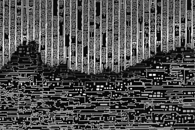
Evgenii Modin's "Microprocessor hieroglyphs", selected photo in FOTCIENCIA
The image "Microprocessor hieroglyphs" captured by Evgenii Modin, Electron Microscopy specialist at CIC nanoGUNE, has been selected as one of the best scientific images of 2021 in FOTCIENCIA. Congratulations!

Evgenii Modin is a doctor in Physics currently working as Electron Microscopy specialist at CIC nanoGUNE. With the objective of giving as much visibility as possible to the science and the research capabilities of the Electron Microscopy Laboratory at nanoGUNE, Modin frequently publishes images related to his work through social networks, especially through the account @electronmicrolab on Instragram.
The image selected in FOTCIENCIA, named "Microprocessor hieroglyphs" was taken with a scanning electron microscope (SEM) at nanoGUNE (Thermo Fisher Helios 450s SEM). The description given to the image is the following: Integrated circuits and microprocessors are part of our daily lives. Today they can be found almost everywhere. They are very complex devices consisting of millions of transistors with dimensions in the order of tens of nanometers. The structure of such semiconductor microworlds can be observed using a scanning electron microscope (SEM). This photo shows a section of the circuit where its design appears to be handwritten in hieroglyphics. The size of these "hieroglyphs" is about 1 micrometer. Using the electron microscope we can not only see the schematic, but we can examine a single transistor to find the defect or improve the technology.
"As a test sample for the alignment of the equipment we used a microprocessor, and we took this opportunity to capture some nice images of the internal structure of the microchip", explains Dr. Modin. That was not something simple, because it requires very precise sample preparation and accurate imaging at high magnification. Some elements of the structure can be very small, well below 1 micrometer (which is a thousand times smaller than a millimeter).
"I have been working in the field of electron microscopy since 2007. I started in Russia (Vladivostok) where I got my PhD in physics and now I am a member of the scientific team of nanoGUNE, where I am a specialist in electron microscopy. So, as a scientist, I am completely immersed in microphotography, and besides, photography is also one of my passions: I really like landscape photography and nightscapes.

